


Eye Adaptation & Fatigue: Optimal Dental Treatment Room Lighting
Introduction
Dental professionals often work under extreme lighting variations – for example, shifting gaze from a moderately bright operatory (~1,500–2,000 lux ambient) to the intense beam of a surgical lamp (~20,000 lux). Such transitions challenge the visual system’s ability to adapt and can contribute to eye strain and fatigue. Understanding how the eye and brain adjust to different light levels is crucial for managing visual comfort in clinical settings. This article explains the mechanisms of visual adaptation (photopic vs. scotopic vision) and how rapid lighting changes induce visual fatigue. It also compares common illuminance levels in various environments (from dim hallways to sunlight), discusses why 2,000 lux feels glaring indoors but not outdoors, and explores how full-spectrum daylight-like lighting can reduce fatigue and support visual performance and circadian health. All claims are grounded in scientific, peer-reviewed research relevant to dentistry and occupational ergonomics.
How the Eye Adapts to Different Light Levels
Human vision operates over an enormous range of illumination – from starlight to bright sunlight – a range of about 10^9 in intensity. To cope with this, the eye adjusts both optically (via the pupil) and photochemically (via retinal photoreceptors and neural adaptation). The pupillary light reflex provides a quick first response: the iris constricts the pupil in bright light and dilates it in dim light, altering retinal illumination. However, the pupil’s effect is relatively minor – roughly a 10-fold change in light intake – which is only a small fraction of the eye’s total adaptive range (en.wikipedia.org).
Figure 1 illustrates this reflex: in bright conditions the pupil is small, whereas in darkness it expands dramatically.
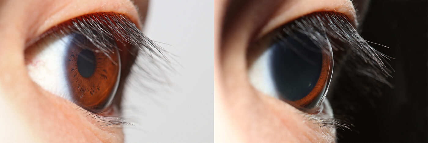
This rapid reflex protects the retina from sudden glare but cannot fully account for the billion-fold range of human vision. Instead, most adaptation occurs in the retina itself, through the dynamic responsiveness of rods and cones.
Figure 1: The pupillary light reflex in an eye exposed to bright light (left) vs. darkness (right). The pupil constricts in bright conditions and dilates in dim conditions as an immediate adaptation, but this mechanism only accounts for about a 10× change in light entering the eye (en.wikipedia.org). Deeper adaptation to ambient light levels is mediated by the photoreceptors (rods and cones) in the retina.
Retinal photoreceptors adjust their sensitivity depending on ambient luminance. In bright environments, cone cells (responsible for photopic vision) dominate. Cones function in high light levels, provide color vision and high acuity, and they recover sensitivity relatively quickly after changes in lighting. In dark or dim environments, rod cells (responsible for scotopic vision) take over. Rods are extremely sensitive to low light and enable night vision (monochromatic and low acuity), but they require more time to fully adapt. The transition between cone-mediated and rod-mediated vision is continuous, defining a mesopic range where both systems contribute (en.wikipedia.org). In numeric terms, above about 0.03 cd/m^2 (a luminance unit), cones chiefly mediate vision (photopic range), whereas below that threshold rods dominate (scotopic range) (en.wikipedia.org). This dual-system design (known as the duplicity theory) allows the visual system to function across vastly different light intensities by switching between cones and rods as needed.
Photopic vs. Scotopic Adaptation Mechanisms
Under bright light (photopic) conditions, cone photopigments bleach and regenerate rapidly. Cones can adapt within minutes to changes in illumination. Under very low light (scotopic) conditions, rods become the main actors; the rhodopsin in rods regenerates slowly after exposure to light, which is why adapting from bright to dark is prolonged. Dark adaptation (entering a dark environment after bright light) occurs in two phases: an initial fast phase driven by cones, followed by a slower, deeper phase as rods gradually regain sensitivity. Within about 5–10 minutes in darkness, rod sensitivity begins to improve dramatically, supplementing the cones. Full rod-based dark adaptation (maximal sensitivity) can take ~30–40 minutes, after which the retina may become over a million times more sensitive than in daylight. Conversely, light adaptation (going from dark to bright) is much quicker – the cones can suppress rod input and adjust to bright light in on the order of a few minutes (en.wikipedia.org). In practical terms, a dentist stepping out of a dark room into sunlight may feel momentarily dazzled, but the cones and neural circuits in the retina rapidly desensitize to bring vision back to comfort within moments. The eye’s neural pathways also contribute to adaptation: retinal neurons and those in the visual cortex adjust their gain and processing based on the average luminance level over time, helping “recalibrate” vision to the prevailing light level.
In summary, rods and cones serve complementary roles in adaptation. Cones mediate vision in well-lit settings and recover quickly, whereas rods enable low-light vision but recover slowly after bright exposure. The retina regenerates photopigments (opsins) and modulates photoreceptor responsiveness as ambient light changes, while the pupil provides a fast but limited adjustment. Together, these mechanisms allow the eye to function across a broad range of lighting, but frequent or extreme shifts in light levels (as often encountered in dental work) can tax the adaptation process.
Typical Illuminance Levels: Indoors vs. Outdoors
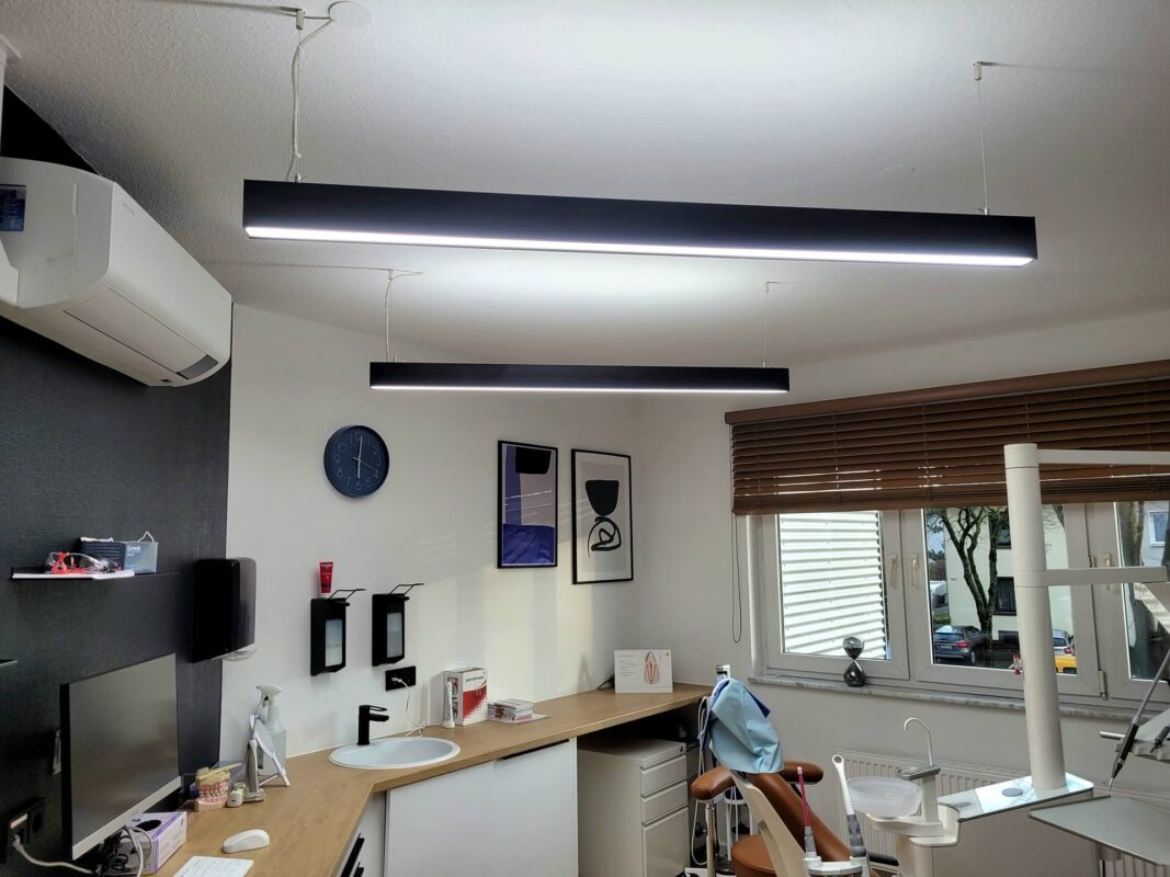
Illuminance (light intensity falling on a surface) is measured in lux. To appreciate the range of lighting conditions the eye can encounter, Table 1 lists typical illuminance levels for various environments, from very dim to extremely bright. Notably, indoor lighting tends to be orders of magnitude lower than outdoor daylight. For context, a dental operatory’s ambient lighting at 2,000 lux is extremely bright by indoor standards, whereas it would be relatively modest compared to outdoor daylight.
Table 1: Typical Illuminance Levels in Various Environments (approximate values in lux) (pmc.ncbi.nlm.nih.gov)
| Environment | Illuminance (lux) |
|---|---|
| Dim hallway or storage area | ~50 lux |
| Living room (evening) | ~300 lux |
| Office or dental operatory ambient | ~500–2,000 lux |
| Overcast outdoor daylight | ~1,000 lux |
| Dental task light (surgical lamp) | ~20,000 lux (at subject) |
| Direct sunlight (midday) | >100,000 lux |
Source: Typical indoor lighting ranges from tens to a few hundred lux, whereas outdoor daylight ranges from thousands to over 100k lux (pmc.ncbi.nlm.nih.gov). (Dental operatory values are estimated from clinical lighting norms.)
As shown in the table, a normally lit indoor environment like an office is around 300–500 lux, and even a brightly illuminated dental clinic ambient may reach 1,000–2,000 lux. These levels are trivial compared to natural outdoor light – a completely overcast sky might still deliver on the order of 1,000 lux, and direct noon sunlight can exceed 100,000 lux (pmc.ncbi.nlm.nih.gov). In essence, a “bright” indoor setting is still dim relative to outdoors. Humans evolved under daylight conditions, and our visual system is adapted to handle very high luminance, but we spend most of our time in much dimmer indoor lighting. This contrast sets the stage for why certain intensities feel glaring in one context but not another, as discussed next.
Why 2,000 lux Feels Bright Indoors but Not Outdoors
An illuminance of 2,000 lux – roughly the brightness of a well-lit dental operatory – can feel uncomfortably intense in an indoor setting, yet the same 2,000 lux under natural outdoor conditions might go unnoticed. This discrepancy is due to the relative perception of brightness and the state of adaptation of our eyes. The visual system responds largely to contrasts and ratios rather than absolute light levels (mdpi.com). Indoors, if the general ambient is a few hundred lux, a localized source of 2,000 lux represents a large jump in luminance, possibly causing glare and discomfort. The pupil may still be relatively dilated (due to the dimmer surroundings), so the bright area delivers a dazzling amount of light to the retina. The sudden high contrast triggers reactions like squinting or “photophobia” (light aversion), a normal response when the eyes are adapted to dimmer light.
Outdoors, by contrast, the overall ambient illumination is usually in the thousands of lux. Under a bright sky, the eyes and brain are pre-adapted to high luminance – pupils are constricted and retinal sensitivity is dampened. In that state, a 2,000 lux stimulus is comparatively small against a background that might be 10,000–20,000 lux or more. The same absolute light level that seemed blinding indoors is just a minor component of the daylight scene, causing little subjective brightness difference. In other words, our brightness perception is relative to the baseline illumination. A given lux level will elicit different sensations depending on one’s current state of adaptation and the surrounding context.
Scientific studies on visual ergonomics reinforce this principle. Discomfort glare occurs when there are large luminance ratios in the field of view – for instance, looking from a dark area toward a bright area. In fact, moving from a darker space to a brighter space induces temporary visual discomfort (sensitivity to light) until adaptation catches up (mdpi.com). One study notes that low-illumination environments “prime” the eyes for high sensitivity, so entering a brighter zone causes sensations of glare or photophobic discomfort until the visual system readjusts (mdpi.com). Thus, 2,000 lux feels glaring indoors precisely because indoor eyes are dark-adapted relative to that level. Outdoors, eyes are light-adapted, so 2,000 lux falls well within the comfort zone.
In dental clinics, this means that if the ambient room lighting is relatively low and then a procedure light of 20,000 lux is switched on, the visual contrast is extreme. The clinician’s eyes, adapted to the lower ambient, will momentarily struggle with the flood of light. If instead the overall operatory lighting is kept higher (though still less than the task light), the relative jump to the spotlight is less severe, reducing perceived glare. Indeed, visual comfort is strongly dependent on maintaining reasonable luminance ratios between task lighting and surrounding areas (mdpi.com). A bright light is more tolerable when one’s surroundings and adaptation level are also on the brighter side.
Visual Fatigue from Frequent Lighting Transitions
Repeated or abrupt changes in light intensity force the eyes to continually adapt, and this process itself can lead to visual fatigue. Visual fatigue (asthenopia) in this context may manifest as eye strain, soreness, difficulty focusing, or headache after long periods of coping with challenging light conditions. In dentistry, a classic scenario is repeatedly glancing from a brightly lit oral cavity under the exam lamp to paperwork or instruments in the comparatively dim periphery, and back again. Each such transition requires the pupil to readjust and the retina to recalibrate sensitivity. Over time, this constant effort can tire the visual system.
Research in occupational lighting and ergonomics confirms that poorly managed lighting transitions can degrade visual performance and comfort. A study on human visual comfort found that “rapid changes in illuminance trigger visual fatigue” and that keeping the illumination levels of adjacent areas closer together (i.e. minimizing large jumps) preserves visual comfort (mdpi.com). In the experiment, subjects’ visual reaction times were used to gauge adaptation; large, sudden increases in illumination caused slower responses and more discomfort compared to more gradual or moderate changes (mdpi.com). The authors concluded that the smaller the difference in illuminance between zones, the more visual acuity is maintained and “visual fatigue can also be avoided.”(mdpi.com). In practical terms, eyestrain was lowest when the lighting contrast was controlled and not extreme.
Medical literature also notes that going from a dark state to a bright state induces temporary visual disturbance. One report states that “walking from a darker space to a brighter space” leads the eyes to experience discomfort (like light sensitivity) until they adapt (mdpi.com). If such transitions happen repeatedly (as with a dentist’s frequent glances between a shadowed area and a lit target), the cumulative stress on the adaptation mechanisms can result in sustained fatigue. Pupil muscles continuously contracting and relaxing, and photoreceptors constantly bleaching and regenerating, create a metabolic and neural load on the visual system. Over a long procedure, this can reduce a clinician’s visual clarity and contribute to general fatigue.
Other factors may exacerbate the strain: if the bright task light also produces glare (e.g., reflecting off an instrument or tooth surface), it further stresses the eye by introducing veiling luminance and reducing contrast. Glare and high luminance ratios have been linked to symptoms of visual strain and even neck/shoulder strain (as the person may adopt awkward postures to avoid glare) in workplace studies (mdpi.com). Thus, managing the transitions and contrasts in lighting is an important part of ergonomics in dentistry. Approaches like using adjustable intensity for the exam light, improving ambient lighting, or allowing a moment for eyes to adjust when looking up from the task can all help mitigate fatigue.
Benefits of Full-Spectrum Daylight Lighting
One strategy to enhance visual comfort in the clinic is the use of full-spectrum (daylight-simulating) lighting in the ambient environment. “Full-spectrum” generally refers to light sources that emit a balance of wavelengths across the visible range, closely resembling natural daylight (including a component of short-wavelength blue light). These lights typically have a high Color Rendering Index (CRI), making colors appear more true-to-life – a clear benefit for dental work that involves color matching of teeth and tissue assessment. But beyond color accuracy, full-spectrum lighting can positively affect visual fatigue and overall well-being.
Daylight-spectrum lighting supports the eye’s natural adaptive expectations. Humans evolved with sunlight as the primary light source, so our visual and circadian systems are tuned to it. Research shows that people subjectively prefer natural light and that it provides measurable health and wellness advantages compared to dull or narrow-spectrum illumination (mdpi.com). For example, exposure to light enriched in the blue spectrum (as in natural daylight) during daytime has been found to improve alertness and cognitive performance (pmc.ncbi.nlm.nih.gov). Short-wavelength (blue) light drives the response of intrinsically photosensitive retinal ganglion cells (ipRGCs) in the eye, which signal to the brain’s circadian clock and acute alertness pathways. IpRGCs have peak sensitivity around 480 nm (blue-turquoise light) (pmc.ncbi.nlm.nih.gov). Thus, a full-spectrum or blue-enriched white light can stimulate these cells effectively, leading to stronger “non-visual” responses like heightened alertness, better mood, and regulated sleep-wake rhythms. Studies have confirmed that, “higher illuminance and short-wavelength (blue) enriched light” elicit greater physiological and alerting responses than dimmer, yellowish light (pmc.ncbi.nlm.nih.gov). In practical terms, a bright, daylight-like clinic environment during working hours can help clinicians feel more awake and potentially reduce the sense of eye fatigue, especially in contrast to working under dim, yellow lighting.
Full-spectrum lighting may also reduce fatigue by improving visual clarity. High CRI daylight lamps ensure that contrast and color differences are sharp, which means the eyes don’t have to work as hard to distinguish details. If a dentist can more easily see fine detail under well-balanced lighting, the visual system experiences less strain. One occupational study found that supplemental task lighting with a broad spectrum improved participants’ visual performance and reduced self-reported eye strain (mdpi.com) (pmc.ncbi.nlm.nih.gov). This aligns with the notion that proper lighting can alleviate the load on the eyes.
Another benefit is the support of circadian health. Clinical staff often work indoors for long hours. Lack of daylight cues can disrupt the normal circadian rhythm, potentially leading to sleep disturbances and fatigue. Providing bright, daytime-spectrum light in the clinic during the day helps reinforce circadian signals – essentially reminding the body that it is daytime, which can improve daytime alertness and later nighttime sleep quality (pmc.ncbi.nlm.nih.gov). Indeed, exposure to “bright light (>1000 lux)” during the day has been linked to better subsequent sleep and alignment of the sleep-wake cycle in several studies (pmc.ncbi.nlm.nih.gov). By contrast, spending the day under dim artificial light (common in many buildings) can leave one feeling sluggish and can blunt the normal circadian contrast between day and night. Full-spectrum lighting, used appropriately, can counteract this by delivering daytime brightness cues. Clinical investigations into light therapy show that bright daylight-mimicking light can reduce stress and improve mood – for instance, it is an established treatment for seasonal affective disorder and has general antidepressant effects(mdpi.com). Even for those not suffering from mood disorders, there is evidence that a pleasant light environment (close to natural light) reduces self-reported stress and tiredness at work (mdpi.com).
In summary, incorporating full-spectrum daylight-like lighting in dental operatories can yield multiple advantages: it lowers visual strain by providing excellent visibility and balanced spectral content, it helps maintain the practitioners’ alertness and circadian rhythm (thus fighting fatigue), and it enhances the overall comfort and mood in the workspace. For example, replacing a dull, flickering fluorescent overhead light with an LED fixture that simulates natural daylight at around 5,500 K color temperature and provides ~1,000–2,000 lux on surfaces can make a noticeable difference. A well-lit operatory with high-quality light allows the eyes to operate in a more “natural” state with less continuous stress. Over a long workday, this can translate to less eye fatigue and possibly even improved precision in visual tasks.
Conclusion
Frequent transitions between vastly different lighting levels, as often experienced in dental practice, pose a challenge to the visual system and can lead to practitioner fatigue. The human eye adapts to light through a combination of rapid pupil adjustments and slower retinal mechanisms involving rods and cones. When these adaptations are pushed to their limits by extreme illuminance contrasts (e.g., 2,000 lux to 20,000 lux back and forth), visual comfort deteriorates. Scientific evidence confirms that large jumps in illuminance induce eye strain and slow visual responsiveness (mdpi.com). On the other hand, maintaining a more consistent lighting environment and employing high-quality, full-spectrum lighting can help the eyes work more comfortably. Dental professionals can benefit from operatory lighting designs that minimize harsh contrasts – for instance, by increasing ambient light levels before activating the operatory lamp, using adjustable intensity headlights, and positioning lights to avoid direct glare. Additionally, using daylight-mimicking lights for general illumination supports the visual system’s natural preferences and the body’s circadian needs, promoting alertness and reducing fatigue (pmc.ncbi.nlm.nih.gov). By understanding and applying these principles, clinicians can protect their vision, reduce eye strain, and maintain better concentration and comfort throughout the workday.
References: (Scientific literature cited)
Citations: Adaptation (eye) – Wikipedia